Case: 6.A.DDD.SL.7.19.20.25
47 Jears (female)
Anamnesis:
Over the last 7-8 years patient complains about pains in the lumbar area while walking, turning and getting up from sitting. Shooting pains with girdle radiating pain into both legs, especially into the left leg. Pains have a circular character. Aching pains are constant in the lumbar area. Feet numbness on the outside, especially on a left. In the last two years the situation worsened.
Diagnosis:
- degeneration and circular protrusion of L3/L4 discs
- deforming degeneration of L4/L5, L4/S1 discs
- spondylolisthesis with a ventralisation of L4 vertebral body
- circular stenosing central foraminal protrusion L4/L5 which caused the spondylolisthesis
- complete stenosis of L5/S1 left foramina
- segment destabilization of L4/L5, L5/S1
- double-sided facet joint arthrosis of L3/L5, L4/L5, L5/S1
Surgical report dated 29.02.2016:
- Intradiscal and intraarticular transplantation of regenerative substances and growth factors concentrate via MIBRAR® method,
- received that was created from venous blood via CGF technology,
- also autologous concentrate from abdominal fatty tissue,
- into the L4/L5, L5/S1 segments.
Surgical report dated 20.04.2017:
- Intradiscal and intraarticular transplantation of regenerative substances and growth factors concentrate via MIBRAR® method,
- received that was created from venous blood via CGF technology,
- also autologous concentrate from abdominal fatty tissue,
- into the L4/L5, L5/S1 segments.
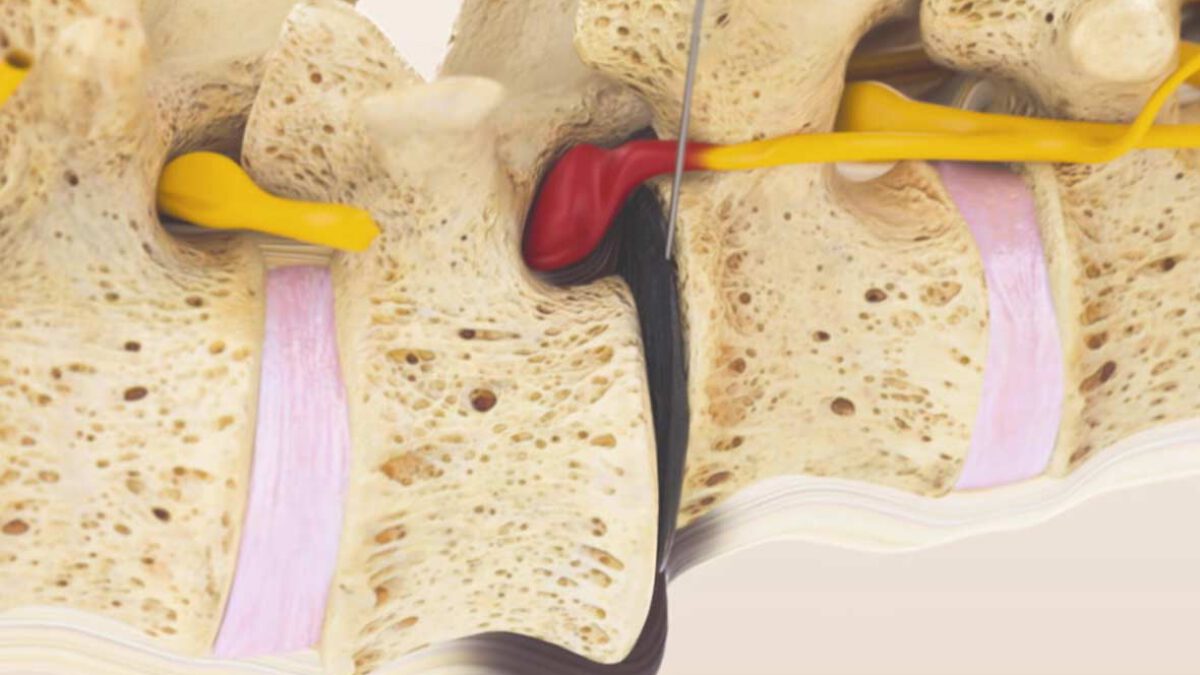
Images description: MIBRAR® surgery of the intervertebral disc L5/S1
Image 1:
Intraoperative sagittal X-ray image dated February, 29 2016 is taken prior to surgery on the spinal disc L5/S1 and corresponds to the preoperative status.
Image 2:
Intraoperative sagittal X-ray image dated February, 29 2016 is taken after the implantation of an autologous CGF concentrate and adipose-derived stem into the spinal disc L5/S1; a massive increase of the disc height can be clearly seen.

Image 3:
The sagittal X-ray image dated May, 12 2016 is a control image and corresponds to the postoperative status ten weeks after the surgery, the increase of the disc height is well illustrated.
Image 4:
The intraoperative sagittal image dated April, 20 2017 is taken during the second surgery and shows further increase of the disc height in 14 months after the MIBRAR® application.

Image 5:
Intraoperative sagittal X-ray image dated April, 20 2017 is taken after the repeated implantation of the autologous CGF concentrate and adipose-derived stem into the spinal disc L5/S1.

MIBRAR® surgery on the intervertebral disc L4/L5 immediately following the surgery on the intervertebral disc L5/S1
Image 6:
The intraoperative frontal X-ray image dated February, 29 2016 is taken prior to spinal sonde surgery on the spinal disc L4/L5 and corresponds to the preoperative status.
Image 7:
The intraoperative frontal X-ray image dated February, 29 2016 is taken after the implantation of an autologous concentrate into the spinal disc L4/L5; a massive increase of the disc height can be clearly seen.

Image 8:
The frontal X-ray image dated July, 8 2016 is a control image and corresponds to the postoperative status 4 months and 10 days after the surgery. It shows that the achieved results of the restoring disc height and form during the surgery Image 7 have been stopped with a minor increase in height only.
Image 9:
The intraoperative frontal X-ray image is taken during the second surgery before the repeated application and shows the surgery result after 14 months. A significant increase of the disc height compared to Image 8 can be clearly identified.

Image 10:
The intraoperative sagittal X-ray image dated February, 29 2016 shows an application of the spinal disc L4/L5 before the implantation of an autologous concentrate; a significant reduction of the disc height and displacement of vertebral body to the front corresponding to the ventrolisthesis diagnosis.
Image 11:
The sagittal X-ray image dated May,12 2016 is a control image and corresponds to the postoperative status in ten months after the surgery; it shows a significant increase of the disc height and a very slight improvement of the displaced vertebral body L4, just a slight improvement of the ventrolisthesis.

Image 12:
The intraoperative sagittal X-ray image dated April, 20 2017 is taken during the second spinal sonde surgery before the implantation of an autologous concentrate; it shows the increase of the disc height compared to the Images 10 and 11, but still just a slight improvement of the ventrolisthesis of the vertebral body L4.
Image 13:
Intraoperative sagittal X-ray image dated April, 20 2017 shows a significant increase of the disc height and improvement of the vertebral body L4 displacement immediately following the implantation of an autologous concentrate.

Image 14:
The sagittal X-ray image dated July, 14 2017 is taken during the follow-up inspection; it shows a complete restoration of the disc height and form of both intervertebral discs, the complete recovery of the vertebral body L4 displacement, ventrolistesis is no longer to be seen.

Surgery results:
Immediately after the first surgery the shooting pains to the left leg noted in previously described anamnesis disappeared. Within 3 days the sensory disturbances were restored. Pains in the lumbar area disappeared within a week. As a result of the surgery the patient was 80 percent pain-free. One month after the second surgery the remaining complaints were remedied.


