Case: 2.A.DDD.7.19.20.25
43 Jears (female)
Current status before the surgery:
Patient complains on girdle pains in lumbar area, radiating to the left leg and hill, also to the right buttock. Kinesalgia. Patient is in need of certain position in seating, standing and any other movements. Clinically visualized forced crocket low back and pelvis position.
Surgery was performed for intervertebral disc hernia in 2013., L5/S1. After what was some temporary relief. But patient started to experience kinesalgia and forced compensatory body position.
Diagnosis:
Status after radiofrequency nucleotomy on lumbar spine.
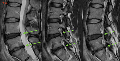
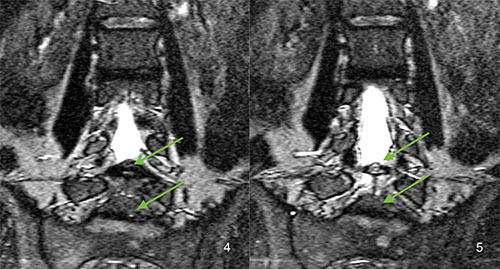
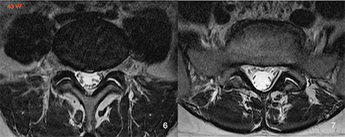
Lordosis changes. Narrowing of intervertebral foramens on L4/L5 and L5/S1 levels. Postoperative disc extrusions of L4/L5 and L5/S1. Activated osteochondrosis on L4/L5, L5/S1 levels. Signs of double sided activated spondiloarthrosis on L4/L5 and L5/S1 levels. Lumbosacral destabilization. L4/L5 disc degeneration and degenerative wear of intervertebral disc L5/S1.
Surgery report:
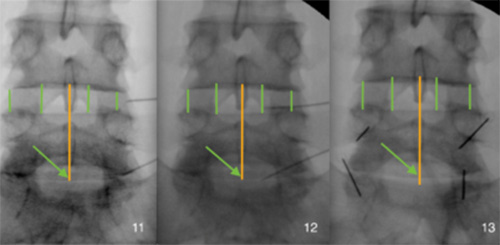
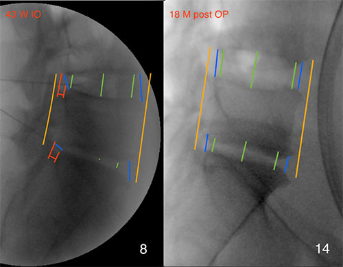
Intradiscal reconstructive implantation of autologous concentrate (composition: blood plasma, young thrombocytes with growth factors, late thrombocytes with growth factors, anti-inflammatory factors) with height and volume restoration of discs under 3D-roentgen control C-arc in intervertebral discs’ area of L4/L5 and L5/S1, dorsal lateral (2 entrances).
Application on L3/L4, L4/L5, L5/S1 facet joints area levels on right and left sides (6 entrances). Peridural implantation of autologous concentrate in lumbar area of L4/L5 and L5/S1 on right and left sides (4 entrances).
Surgery result:
All the symptoms disappeared in a week after the surgery.
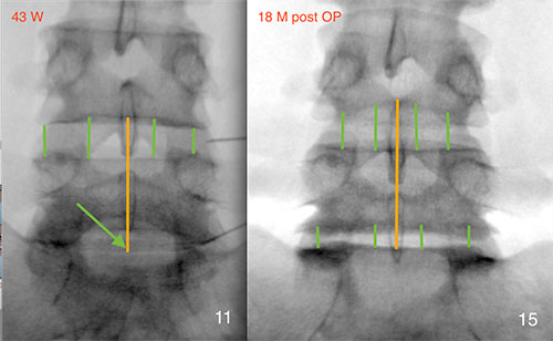
Below are the images of intraoperative roentgen before and after reconstructive implantation, and also MRI images before the surgery. On intraoperative roentgen images very well visualized the height and volume restoration and restoration of intervertebral disc shapes.
MRI images description:
Above MRI images before the surgery; below intraoperative roentgen images